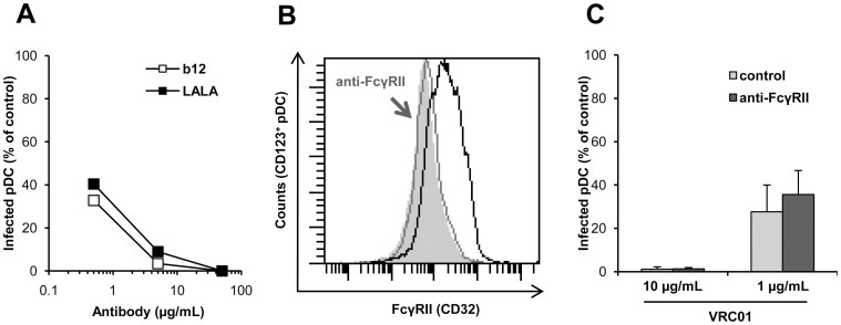
Figure 3. Role of FcγRII in the inhibition of infection of primary pDC by HIV-1.
(A) Data represent the percentage of pDC infected by HIV-1BaL in the presence of various concentrations of NAb b12 or LALA (µg/ml) compared to the control condition without NAb. Data are representative of three independent experiments with primary pDC from three different healthy donors. (B) Detection of FcγRII on pDC (black line) or after 30 minutes of incubation with anti-FcγRII Ab (10 µg/ml) (grey line, arrowhead); isotype control in grey. (C) pDC were pre-incubated with or without anti-FcγRII Ab (10 µg/ml) for 30 minutes at 37°C before being added to the HIV-1BaL/VRC01 mix. Percentage of infected cells in the presence of NAb VRC01 compared to the control of infected cells in the absence of NAb is shown. Data are means ± SD of two independent experiments performed with primary pDC purified from blood of two healthy donors.

