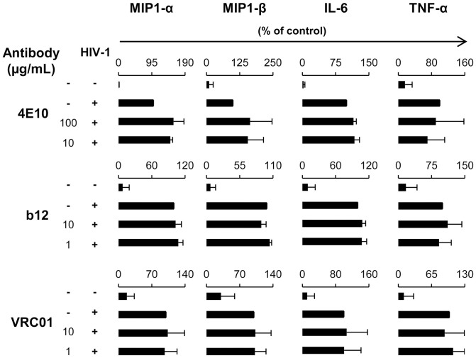
Figure 6. Production of MIP1-α, MIP1-β, IL-6 and TNFα by primary pDC during neutralization assay.
Supernatants of pDC were collected after 48 h of infection and screened by CBA Flex to measure MIP1-α, MIP1-β, IL-6 and TNFα. The concentration of MIP1-α, MIP1-β, IL-6 and TNFα in the supernatants from pDC incubated alone (Mock) or with HIV-1BaL (HIV) or with HIV-1BaL in the presence of NAb 4E10, b12 or VRC01 is shown. Data are representative of three independent experiments performed with primary pDC from three healthy blood donors.

