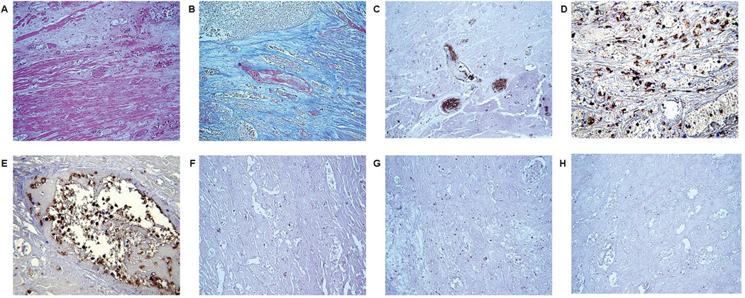
Figure 2.
Histopathologic features of the heart graft that failed after 8 weeks (Exp 12), showing (A) thrombosis and infarction (H&E, x200), (B) fibrin deposition (MSB x400), and (C) massive platelet accumulation (IHC, x400). A neutrophil infiltrate can be seen in the interstitium (IHC, x400) (D); these cells are also present in the vessels of the graft (E), but there are relatively few infiltrating macrophages (IHC,200) (F), and very few T (G) and B (H) lymphocytes (stained brown) (magnification x200).

