Figure 3.
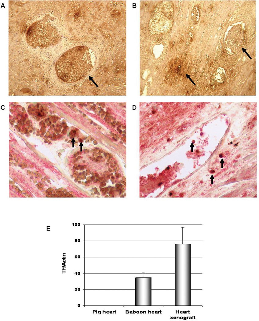
Staining for primate TF in heart grafts that rejected at day 12 (A; Exp 5) and at 8 weeks (B; Exp 12), showing strong staining in the thrombosed vessels and less staining in the interstitium (arrows) (x600). Co-localization of primate TF (red stain) and macrophages (stained for CD68, brown) in heart grafts excised on day 12 (C, Exp 5) and at 8 weeks (D, Exp 12) is indicated by arrows (x600). Biopsies from 3 other rejected xenografts showed similar results. Primate TF mRNA levels in a rejected porcine heart xenograft by qRT-PCR (E); the heart failed from TM at 8 weeks (Exp 12) and showed high levels of primate TF. Tissue from a normal baboon heart was used as a positive control, while a porcine heart was used as a negative control to ensure the specificity of the primate TF primer. Real-time PCR data were plotted as the ΔRn fluorescence signal versus the cycle number. The expression of each gene was normalized to actin mRNA content and calculated relative to control using the comparative CT method.

