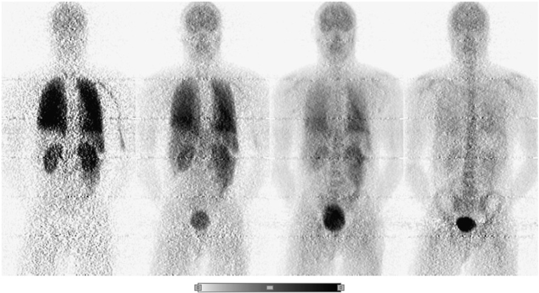
FIGURE 1.
Example of a series of whole-body 2D planar PET images for one healthy male. Images were obtained approximately 2, 20, 100, and 240 min after intravenous injection of about 192 ± 7 MBq (5.2 ± 0.2 mCi) of [18F]SPA-RQ. All 4 decay-corrected images used the same color scale (bottom of figure).

