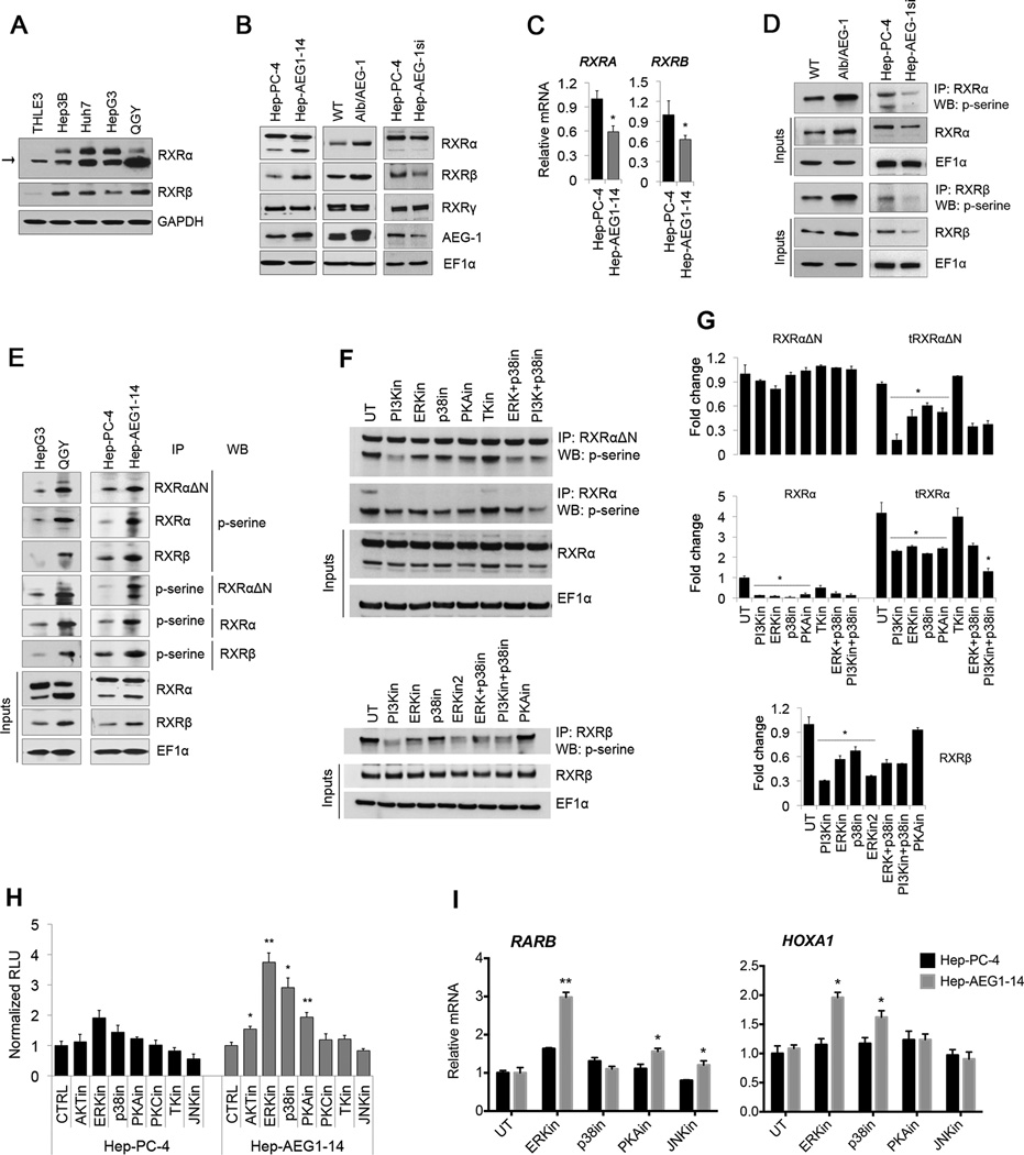
Fig. 5. AEG-1 induces RXR phosphorylation by activating ERK, p38MAPK and PKA signaling.
(A) Expression analysis of RXRα and RXRβ in different HCC cell lines and human immortalized hepatocytes THLE3 cells.
(B–C) Expression of RXRα, RXRβ, RXRγ and AEG-1 in human Hep-PC-4 vs. Hep-AEG1–14 cells and WT vs. Alb/AEG-1 mice hepatocytes at protein level detected with western blot (B) and at mRNA levels by qPCR (C).
(D–E) Phosphorylation levels of RXRα and RXRβ were determined by immunoprecipitating cell lysates with anti-RXRΔN, anti-RXRα and anti-RXRβ antibodies followed by WB with anti-phospho-serine (p-serine) antibody and vice-versa, for WT vs. Alb/AEG-1 mice hepatocytes, Hep-PC4 vs. Hep-AEG-1si (D), HepG3 vs. QGY-7703, and PC-4 vs. AEG1–14 cells (E).
(F–G) Decreased levels of phosphorylated RXRα and RXRβ in Hep-AEG1–14 cells after treatment with inhibitors of the indicated kinases. IP was performed with anti-RXRΔN and anti-RXRα and anti-RXRβ antibodies and WB was performed for p-serine. Graphical representations of densitometry quantification are shown in G. Cells were treated with different inhibitors in DMEM containing 1% charcoal stripped FBS for 24 hours. For concentrations used and detailed information, please see Table S2.
(H–I) RARE activity (H), expression of representative RXR/RAR target genes, RARB and HOXA1 (I) in Hep-PC-4 and Hep-AEG1–14 cells after inhibition of different kinases.
For F–I, selective inhibitors for PI3K, ERK, JNK, TK were used at final 15µM concentration and PKA, PKC and p38 MAPK at 2 µM. Data represents mean ± SEM of three independent experiments. *: p<0.02 and **: p<0.001.

