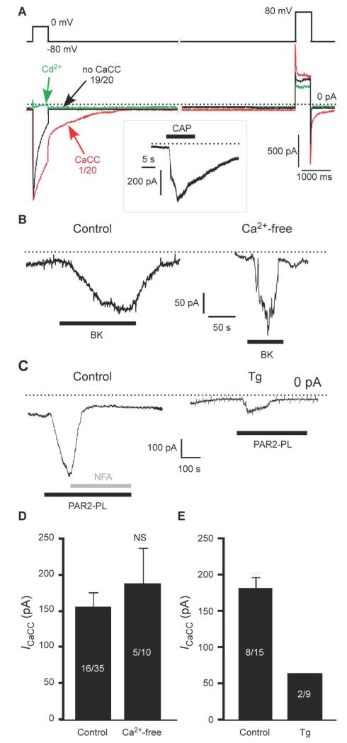
Fig. 1. Ca2+-activated Cl− channels in small DRG neurons are preferentially activated by Ca2+ release from the ER.
(A) Whole cell patch-clamp recording of currents in response to a voltage pulse from −80 to 0 mV in small DRG neurons. The recordings were made using TEACl-based extracellular and CsCl-based intracellular solutions. The green trace represents inhibition of VGCC by Cd2+ (100 μM) in the same cell in which the control (black) trace was recorded. The box inset depicts the response of the same cell to capsaicin (CAP; 1 μM) at a holding potential of −60 mV. 19/20 and 1/20 represent the number of neurons exhibiting the specified type of response. (B) BK (1 μM)-induced inward current in a small DRG neuron when recorded in extracellular solution with and without Ca2+. (C) PAR2-PL (10 μM)-induced inward current in a small DRG neuron; in control conditions or following pretreatment with thapsigargin (Tg, 2 μM, 3 min); NFA, niflumic acid (100 μM). (D, E) Summary data for (B) and (C), respectively. Number of responsive neurons out of total neurons tested is indicated within each bar.

