Figure 1.

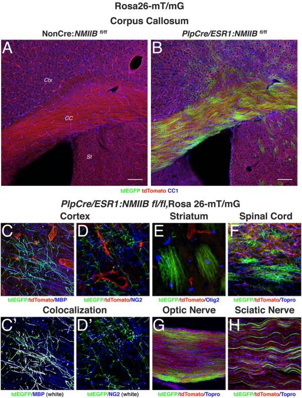
Analysis of tamoxifen-driven PlpCre/ESR1 recombination. Rosa26-mT/mG mice have loxP sites on either side of a membrane-targeted tdTomato cassette and express strong red fluorescence in all tissues. When bred to PlpCre/ESR1 mice, the resulting offspring have the mT cassette deleted in the Cre-expressing cells, allowing expression of the membrane-targeted EGFP cassette. NMIIB fl/fl mice carrying both the PlpCre/ESR1 transgene and the Rosa26-mT/mG allele (Plp-Cre+) or Rosa26-mT/mG allele alone (NonCre) were injected with tamoxifen for 5 days at 8 weeks of age. The animals were sacrificed at 12 weeks and their tissue was processed for immunofluorescence with antibodies to CC1, MBP, NG2, Olig2, and the nuclear stain Topro3. (A, B): Sections of the corpus callosum (CC) show high EGFP expression in OLs in the white matter tract as well as in the cerebral cortex (CTX) and striatum (St). Scale bars 100 µm. (C–H): Details of cells expressing EGFP throughout the CNS. In the cortex (C, D) EGFP label colocalizes (shown in white) with mature OL markers such as MBP (C′), but not with NG2 (D′). Recombination is also observed in myelinating OL in the striatum (E), spinal cord (F) and optic nerve (G) as well as in myelinating Schwann cells in the sciatic nerve (H).
