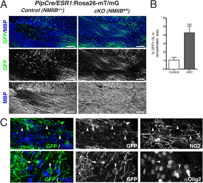
Figure 7.

Myelinating oligodendrocytes lacking NMIIB are present in remyelinated areas. (A): Representative images from 28 dpi lysolecithin lesions from transgenic PlpCre/ESR1:Rosa26-mT/mG mice carrying wild-type (Control) or floxed (cKO) NMIIB alleles. EGFP+ myelinating cells within the remyelinated MBP+ area (blue) were more frequently found in cKO animals. Scale bar 50 µm. (B): Quantitation of percentage area remyelinated by EGFP+ OLs in control and cKO animals (Mann Whitney t-test ***P < 0.001). Data collected from three animals per genotype, three to five fields per animal. Scale bars 50 µm. (C): Examples of NG2− (top panel); Olig2+ (bottom panel) EGFP-expressing OL (arrowheads) found in the lesion border of remyelinated areas in double transgenic cKO mice.
