Figure 1.
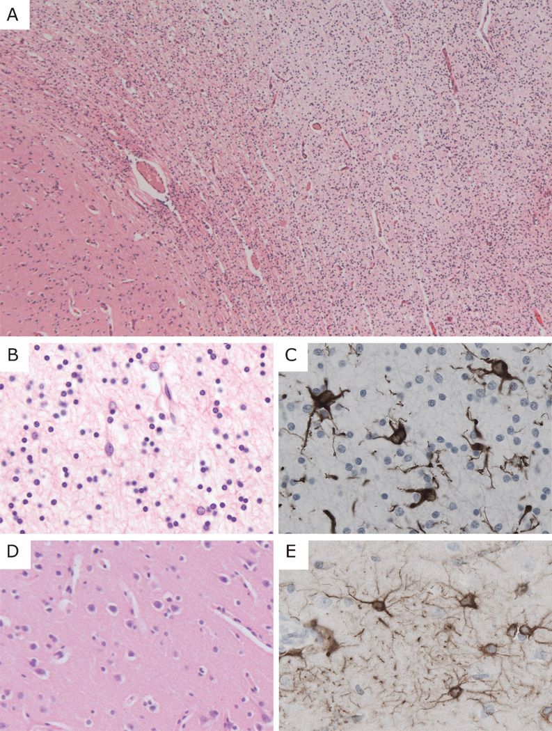
General neuropathological features of vanishing white matter disease. (A) Low-power magnification hematoxylin and eosin staining of the white matter (right side of field) from the frontal lobe of patient VWM367 shows tissue rarefaction and increased cellular density. The U-fibers appear relatively preserved; the overlying cortex is normal. (B, C) At higher magnification, white matter astrocytes display abnormal morphology with coarse blunt processes. (D, E) The cortical architecture and morphology of cortical astrocytes are normal. (A, B, D) Hematoxylin and eosin; (C, E) immunohistochemistry for glial fibrillary acidic protein. Original magnifications: A, 25×; B–E, 200×.

