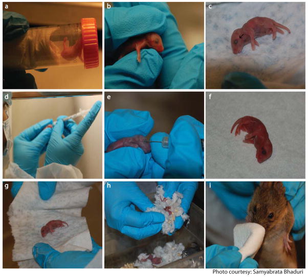
Figure 3.
Intravenous delivery of rAAVs to neonatal mice. Mouse pups are anesthetized in the makeshift anesthesia chamber containing isofluorane (a). Pups are positioned to expose the superficial temporal vein (b). Black arrow indicates the vein (c). Dosing syringe is positioned in dominant hand (d). Needle is slowly inserted into the vein and plunger is depressed (e). Blood flow is stemmed and appearance of a hematoma below the eye indicates successful injection (f). Pups are rolled in alcohol soaked tissue (g) followed by rubbing with dirty bedding (h) before putting them back in the cage. Dam is nose numbed with alcohol prior to introduction into the cage (i).

