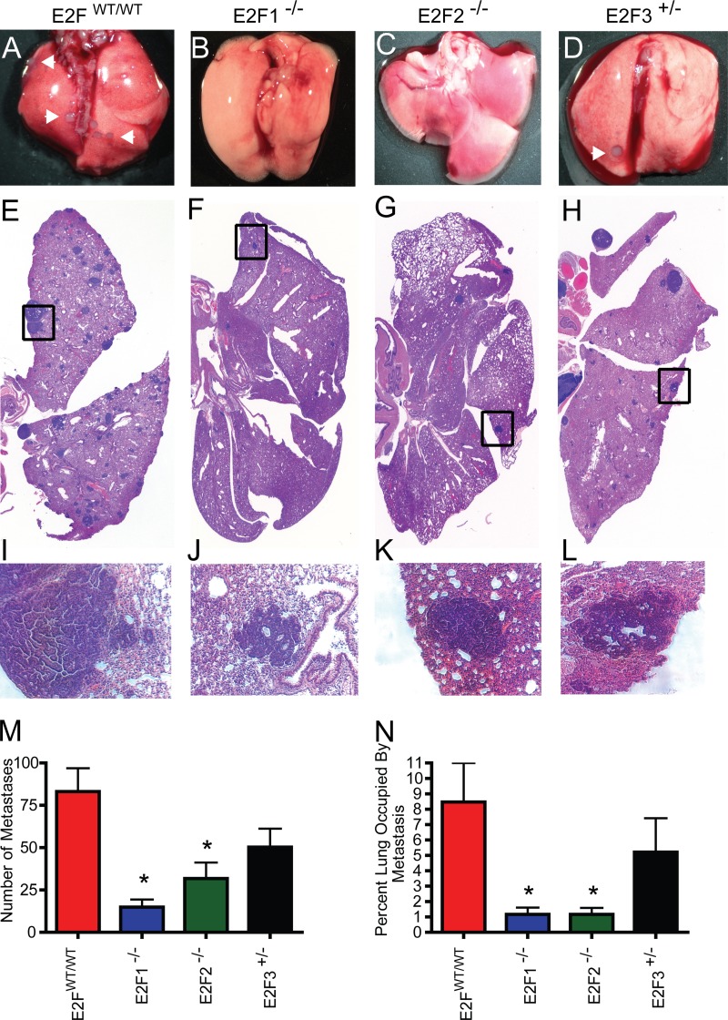
FIG 5.
Loss of E2Fs decreases the level of pulmonary metastasis in MMTV-PyMT mice. (A to D) Representative wet-mount images of lungs from E2FWT/WT, E2F1−/−, E2F2−/−, and E2F3+/− mice. Arrowheads indicate surface metastases. (E to H) Representative images for H&E-stained sections of lungs. (I to L) Tenfold-enlarged images of the regions boxed in panels E to H show the histology of tumor metastases. (M) Comparison of the average numbers of lung metastases (with standard errors of the means) in pulmonary sections from E2FWT/WT mice with those for mice in E2F mutant backgrounds revealed significant differences in E2F1−/− (P, <0.0001) and E2F2−/− (P, 0.002) mice. (N) Comparison of the average percentage (with standard errors of the means) of the lung area occupied by metastases in pulmonary sections from E2FWT/WT mice with those for E2F1−/− mice (P, <0.0001), E2F2−/− mice (P, <0.0001), and E2F3+/− mice.

