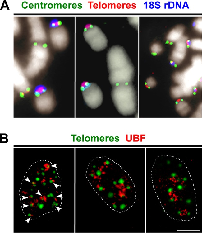
FIG 1.

Position of centromeres and nucleolar relationship of acrocentric chromosomes in primary S. granarius fibroblasts. (A) Centromere position on S. granarius acrocentric chromosomes. A two-step experiment was performed, including immunofluorescence assay with ANA-CREST antibodies to detect centromeric proteins (green) and subsequent two-color FISH with a C-rich telomeric PNA probe (Cy3; red) and an 18S rDNA probe (pseudocolored blue). Chromosomes were counterstained with DAPI (pseudocolored white). Centromeres are generally found adjacent to or overlapping signals from telomeric and rDNA probes. (B) Visualization of transcriptionally active nucleoli in primary S. granarius fibroblasts by use of anti-UBF1 antibodies (red). Telomeres were subsequently visualized through PNA FISH (green). In the optical slice at left, arrows indicate close contacts between telomeres and nucleoli. The mean number of nuclear telomere foci (± SEM) was 13.7 ± 2.8 (range, 9 to 26), with 65% ± 2.6% of them being in association with UBF1 signals (n = 90 nuclei). Bar, 10 μm.
