
FIG 2.
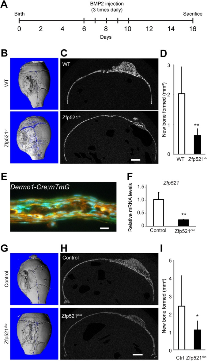
Reduced BMP2-induced bone formation in Zfp521−/− mice. (A) Schematic of a BMP2 calvarial injection model. (B and C) μCT 3D reconstruction (B) and cross-sectional image (C) of wild-type (WT) and Zfp521−/− calvarium following treatment with BMP2. (D) μCT quantification of newly formed bone in response to BMP2. (E) Fluorescence image of a 6-day-old Dermo1-Cre:mT/mG calvarium demonstrating Cre activity (green) in periosteal cells. (F) Expression of Zfp521 in calvaria of 6-day-old Dermo1-Cre:Zfp521f/f (Zfp521cko) mice. (G and H) μCT 3D (G) and cross-sectional (H) images of control (Dermo1-Cre; Zfp521+/+) and Zfp521cko mice following treatment with BMP2. (I) μCT quantification of newly formed bone in response to BMP2. The data are presented as means and SD; n ≥ 3. *, P < 0.05; **, P < 0.01; Student's t test. Scale bars, 1 mm (C and H); 10 μm (E).