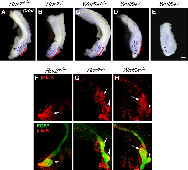
FIG 3.
Gdnf expression is mislocalized in Ror2- and Wnt5a-deficient kidneys. (A to E) Whole-mount in situ hybridization of kidneys (E11.0) with Gdnf antisense probe (brackets). The WD and UBs are outlined. Scale bar, 0.1 mm. (F to H) Whole-mount immunofluorescence staining of kidneys (E11.0) expressing HoxB7-EGFP (green) with anti-p-Erk antibody (red). Images of anti-p-Erk staining alone (top row) and merged images (bottom row) are shown. Arrows indicate UBs. Scale bar, 0.1 mm.

