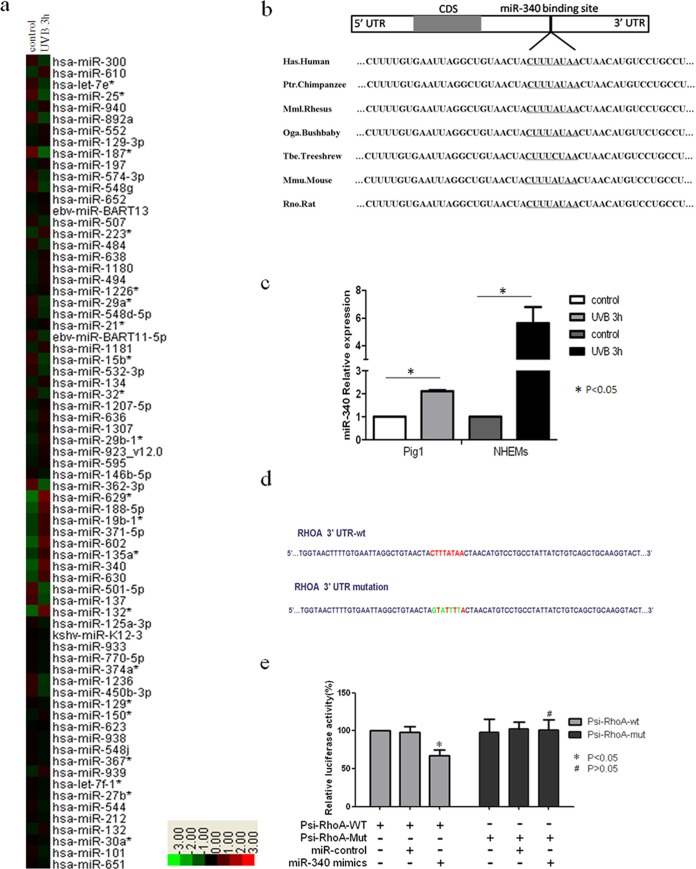
FIG 2.

Identification of miRNAs involved in UVB-induced dendrite formation. (a) Hierarchical clustering analysis of miRNA expression in Pig1 cells without UVB irradiation or 3 h after UVB treatment. Red, +1.5-fold change in gene expression (high expression); green, −1.5-fold change in gene expression (low expression). (b) miR-340 target binding site in RhoA, which was predicted as a target of miR-340 using computational methods. The miR-340 seed binding region downstream of the coding sequence (CDS) is located in the 3′UTR of RhoA, and the sequence of this miR-340 seed binding region is identical in different species. (c) qRT-PCR analysis of miR-340 expression 3 h after UVB radiation in Pig1 cells and human epidermal melanocytes (NHEMs). The expression of miR-340 was normalized relative to the expression of U6 snRNA. Expression is presented relative to the controls, as the means ± SEM. *, P < 0.05, Student's t test. (d) Two luciferase reporter constructs that contained either the wild-type RhoA 3′UTR, including the putative binding site for miR-340, or the RhoA 3′UTR containing mutations within the putative targeting region were generated. The wild-type and mutated RhoA 3′UTR sequences are shown. (e) Wild-type and mutated RhoA 3′UTR sequences were inserted into the psiCHECK-2 vector to create Psi-RhoA-wt and Psi-RhoA-mut constructs, respectively. Each of these constructs was transiently cotransfected into Pig1 cells, along with either miR-340 mimics or control miRNA (miR-control), and luciferase activity was then measured 24 h after transfection. Luciferase activity is shown relative to that in Pig1 cells transfected with Psi-RhoA-wt alone, and data are presented as the means ± SEM. *, P < 0.05; #, P > 0.05 (Student's t test). (f) qRT-PCR analysis of primary and mature miR-340 expression UVB radiation in NHEMs. The expression of primary or mature miR-340 was normalized relative to the expression of GAPDH (glyceraldehyde-3-phosphate dehydrogenase) or U6 snRNA. Expression is presented relative to the controls, as the means ± SEM. *, P < 0.05, Student's t test. (g) P53, Drosha, and p68 (DDX5) protein expression in UVB-irradiated and control cells, detected by Western blotting.

