FIG 5.
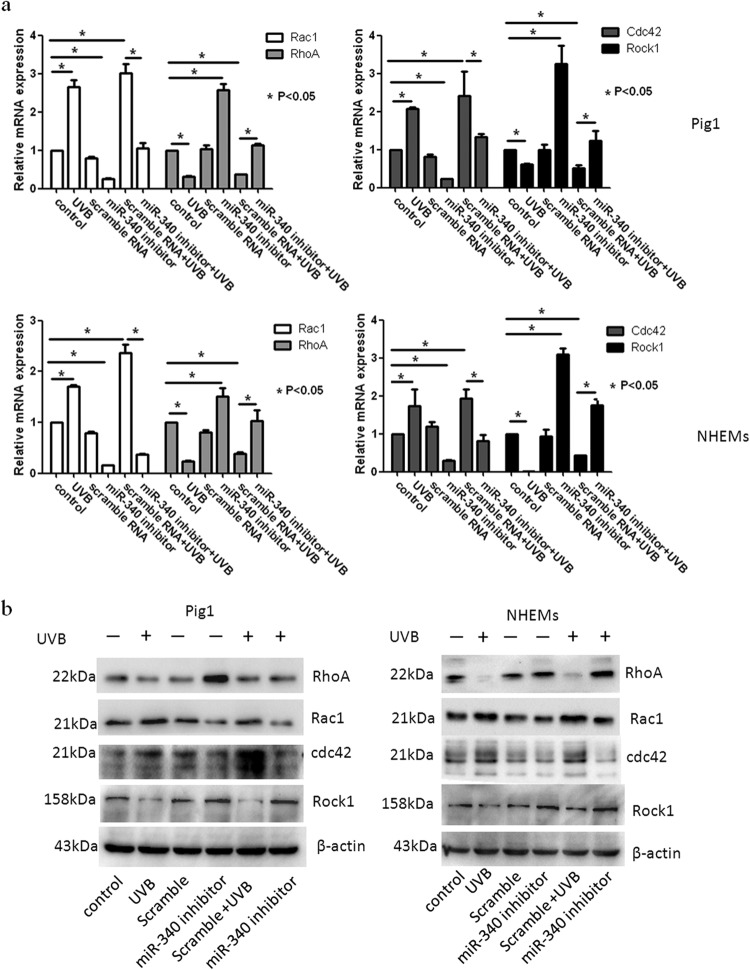
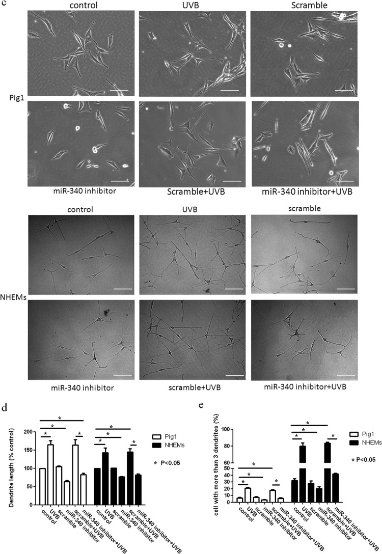
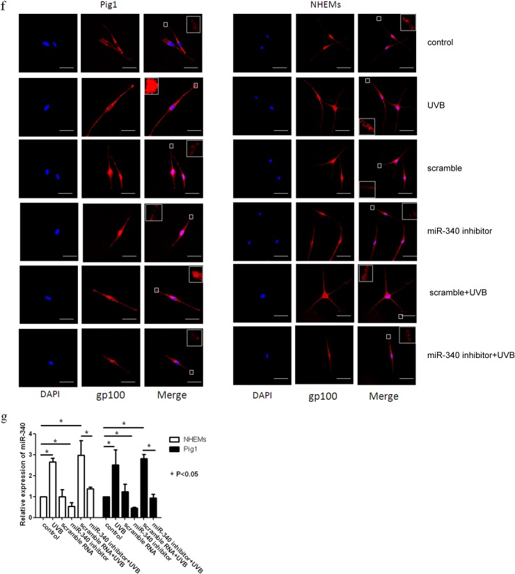
Inhibition of miR-340 blocks UVB-induced dendrite formation. Pig1 cells and NHEMs were exposed to 100 mJ · cm−2 UVB, with or without transfection with miR-340 inhibitors or control (scrambled-sequence) miRNA. (a) Rac1, RhoA, Cdc42, and Rock1 mRNA expression was assessed by qRT-PCR. Expression of mRNA is presented relative to the untreated control, and data are presented as the means ± SEM. *, P < 0.05, Student's t test. (b) Western blotting was used to detect the expression of Rac1, RhoA, Cdc42, and Rock1 proteins. (c) Inhibition of miR-340 blocks UVB-induced morphology change. The morphology of melanocytes was acquired after exposure to 100 mJ · cm−2 UVB, with or without transfection with miR-340 inhibitors or control (scrambled-sequence) miRNA by bright-field microscopy using a 40× objective lens; scale bar, 100 μm. (d) The lengths of cell dendrites were measured by AxioVisionRel 4.8.2 software. Dendrite length is shown relative to that of untreated control cells, and graphs depict the means ± SEM. Student's t test was used to compare dendrite lengths between groups. *, P < 0.05. (e) The dendrites in Pig1 cells from each experiment were manually counted, and sample groups were compared using the Student t test. The percentage of cells with >3 dendrites is shown for each group as the means ± SEM. *, P < 0.05. (f) Inhibition of miR-340 blocks UVB-induced melanosome transport. Pig1 cells and NHEMs were stained with anti-gp100 antibody (red) and DAPI (blue) and then viewed under a confocal microscope using a 60× objective lens to determine melanosome localization within cells. Scale bar, 50 μm. (g) miR-340 expression was assessed by qRT-PCR after exposure to 100 mJ · cm−2 UVB, with or without transfection with miR-340 inhibitors or control (scrambled-sequence) miRNA.


