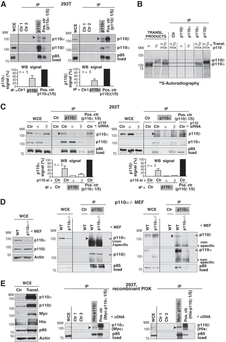
FIG 1.
Endogenous p110α and p110β form a complex. (A) Endogenous p110α or p110β was immunoprecipitated from 293T cell extracts (1 mg) by using specific Ab. WB was used to test for associated p110. A smaller amount (1/5) of whole-cell extract (WCE; 200 μg) was used to immunoprecipitate p110β or p110α positive controls. Negative controls included protein A plus Ab (Ctr 1) and extract plus protein A (Ctr 2); WCE (50 μg) and immunoprecipitates (IP) were analyzed by WB. Graphs show the percentage of p110α signal in the p110β immunoprecipitates and in the p110α positive control, normalized for p85 signal, which estimates the amount of PI3K precipitated under both conditions. Normalized signals are shown as a percentage of maximal signal (p110α positive control, 100%; mean ± standard error of the mean, n = 5). The percentage of p110β in p110α immunoprecipitates was calculated similarly. (B) cDNAs encoding p110α, p110β, or both were transcribed/translated in vitro in the presence of [35S]methionine. Translation products or immunoprecipitated products were visualized by autoradiography. As controls, translation products were incubated with protein A. Frame with solid line, p110α signal; frame with dashed line, p110β signal. (C) 293T cells were transfected with control, p110α, or p110β siRNA (72 h). p110β, p110α, and positive controls were immunoprecipitated from WCE as described for panel A. The negative control contained cell extract plus protein A. The graphs are similar to those for panel A. n = 3. (D) p110αflox/flox MEF were infected with adenoviral Cre (72 h); pik3ca gene deletion was 80% efficient. Cre-infected or WT p110α MEF were lysed and tested as described for panel A. The figure shows a representative experiment (n = 3). (E) 293T cells were transfected with cDNAs encoding Myc-p110α, His-p110β, HA-p85α, and HA-p85β (48 h). WCE (50 μg) was tested by WB or immunoprecipitated (1 mg) using anti-His Ab (for p110β); associated p110α was analyzed with WB. For the positive control, we immunoprecipitated Myc-p110α-transfected cell extract (200 μg) with anti-Myc Ab. Negative controls were as described for panel A; the reciprocal assay was performed similarly. For panels A, C, D, and E, empty lanes rule out spillover from adjacent lanes. Arrowheads indicate the p110 isoform (p110α or p110β) in complex with the other. MW, molecular weight.

