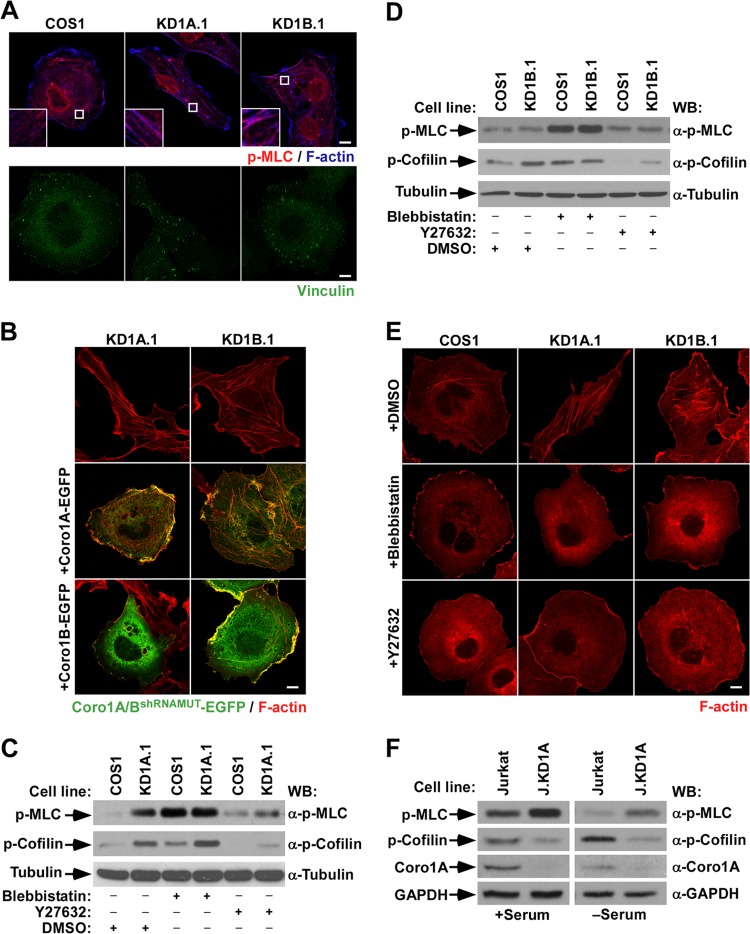
FIG 1.
Unregulated myosin II-dependent contractility contributes to the cytoskeletal phenotype of CORO1 knockdown cells. (A) Representative images of the indicated cells stained with antibodies to phospho-MLC (top row, red signals) and Alexa Fluor 635-labeled phalloidin (top row, blue signals) and with vinculin (bottom row, green signals). In the top row, magnifications of the boxed cell areas are shown in the insets. Color codes for fluorescence signals are indicated. Colocalization of phospho-MLC and F-actin is shown in purple (top row). Scale bars, 10 μm. (B and E) Representative images of the cytoskeletal phenotype exhibited by CORO1 knockdown cells (top) upon ectopic expression of the indicated Coro1-EGFP proteins (B, left) and drug treatments (E, left). In panel B, colocalization of Coro1 and F-actin is shown in yellow. Scale bars, 10 μm. (C, D, and F) Total cellular extracts from the indicated cell lines and experimental conditions were analyzed by Western blotting (WB) to detect the amount of phospho (p)-MLC (top gels), phosphocofilin (second gels from top), and Coro1A (F, third gel from top). The loading controls used were tubulin α (C and D, bottom) and GAPDH (F, bottom). The antibodies used in the immunoblot analyses are indicated on the right.

