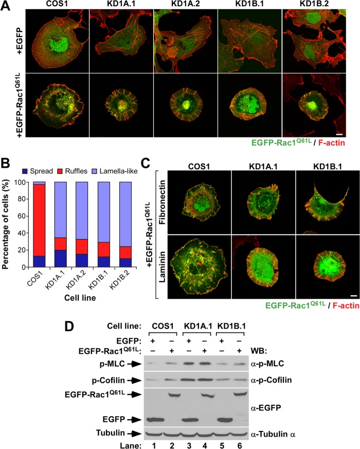
FIG 2.
Abnormal Rac1 cytoskeletal signaling in CORO1 knockdown cells. (A and C) Representative images of the cytoskeletal phenotype exhibited by EGFP-expressing (A) and EGFP-Rac1Q61L-expressing (A and C) cell lines attached to polylysine-coated (A), fibronectin-coated (C), or laminin-coated (C) coverslips. The cell lines are indicated at the top. Ectopically expressed proteins are indicated at the left. Colocalization areas of active Rac1 with F-actin are shown in yellow. Scale bars, 10 μm. (B) Quantification of the types of cytoskeletal change induced by ectopically expressed EGFP-Rac1Q61L in the indicated cells attached to polylysine-coated coverslips. (D) Total cellular extracts from the indicated cell lines and experimental conditions were analyzed by Western blotting to detect the amounts of phospho-MLC, phosphocofilin, and EGFP proteins expressed. Tubulin α was used as a loading control.

