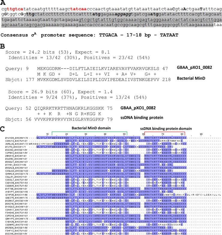
FIG 5.
Nucleotide sequence of minP gene and amino acid sequence of MinP protein in comparison with cognate proteins from B. cereus group. (A) minP nucleotide sequence. The −35 and −10 regions of the σA-like promoter are indicated in red. Five transcriptional start sites identified by transcriptome sequencing (RNA-seq) (29) are shown in uppercase with bold. The structural part of the gene is shaded. Three potential translation start codons are shown in bold, while the consensus Shine-Dalgarno sequence is in italics with bold. The 67-bp stem-loop is underlined. (B) Alignment of MinP using Meta-Server for protein domain searches (http://prodata.swmed.edu/MESSA/). (C) MinP alignment. The Pfam protein database was used to align proteins. The top row shows the Ames Ancestor pXO1 MinP amino acid sequence. Amino acids matching the top reference sequence are indicated in dark violet. Amino acids with similarity to the reference are shown in light violet. The bacterial MinD domain and the ssDNA-binding protein domain are indicated by green and red brackets, respectively.

