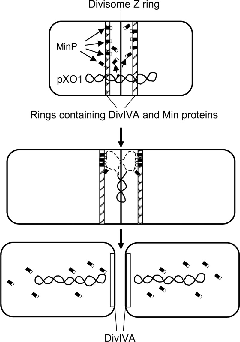
FIG 6.
Model for pXO1 distribution between incipient cells by MinP protein. (Top) The divisome Z ring is flanked by two rings containing DivIVA and Min proteins, modeled after reference 31. The N-terminal part of the MinP protein (indicated as black area in MinP rectangle) is intercalated into the flanking rings. (Middle) Two DNA single-strand loops (indicated by dashed lines) generated during pXO1 replication interact with the C-terminal part of MinP protein (indicated as white area in MinP rectangle). (Bottom) Two pXO1 copies fixed on different rings move to the adjusted poles of dividing cells and, after the cells divide, are released. MinP and other Min proteins leave the rings, and DivIVA reorganizes.

