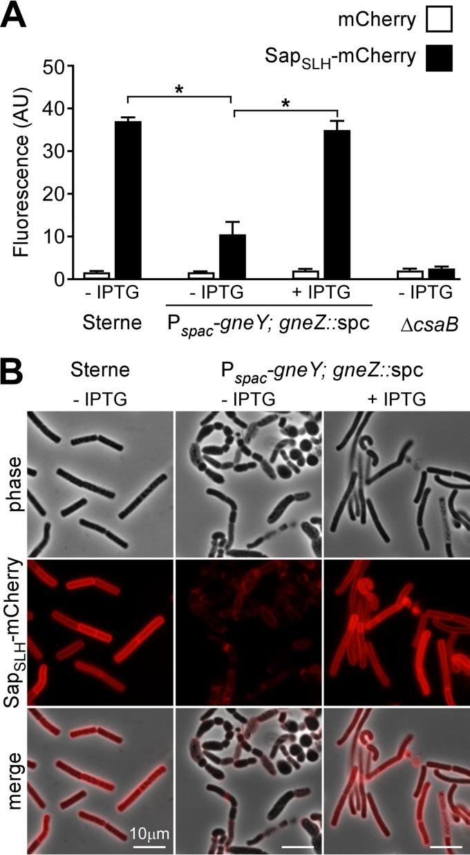
FIG 7.
B. anthracis secondary cell wall polysaccharide synthesis with and without UDP-GlcNAc 2-epimerase expression. Spores derived from B. anthracis Sterne(pJK4) and YW6(pJK4) and the ΔcsaB strain were diluted into BHI without (−IPTG) or with (+IPTG) 1 mM IPTG supplement and incubated for 6 h. The S-layer of vegetative forms was extracted with 3 M urea, and bacilli were incubated with purified mCherry or SapSLH-mCherry. (A) Binding of fluorescent protein (mCherry or SapSLH-mCherry) to bacilli was monitored by analyzing the fluorescence intensity of the bacterial sediment. Fluorescence intensity (arbitrary unit [AU]) measurements were normalized to A600 data, i.e., the bacterial densities of each sample. AU/A600 values were averaged, and statistical significance was examined with the unpaired two-tailed Student t test (*, P < 0.001). (B) Images of samples in panel A were acquired by phase-contrast and fluorescence microscopy. Bars, 10 μm. Data are representative of two independent experimental determinations.

