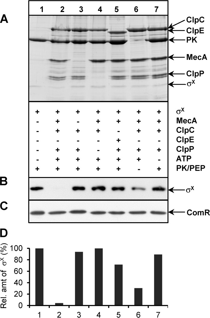
FIG 4.
In vitro degradation of σX. (A) Top, SDS-PAGE with Coomassie blue staining of various combinations of σX-Strep, 6His-MecA, 6His-ClpC, 6His-ClpE, 6His-ClpP, ATP, and the pyruvate kinase/phosphoenolpyruvate (PK/PEP) ATP regeneration system. Bottom, presence (+) or absence (−) of a specific compound in the reaction mixture. (B) σX-Strep detection by Western blotting. (C) ComR-Strep detection by Western blotting. ComR-Strep was used as negative control and substitute for σX-Strep in a parallel experiment performed under the same conditions. (D) Quantifications by densitometry of the relative amount of σX-Strep detected by Western blotting, using control lane 1 as 100%. The specific in vitro degradation of σX in the presence of MecA-ClpCP and ATP has been reproduced with 4 independent batches of purified proteins. The presented experiment with all the controls on one gel was performed in duplicates, which showed similar results.

