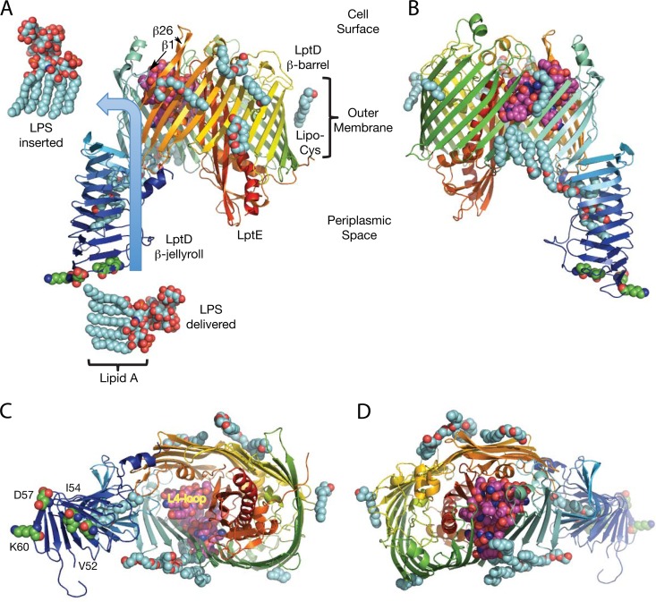
FIG 1.
Structure of the LptDE complex for delivering and inserting LPS into the bacterial outer membrane. (A) The LptDE complex from Shigella flexneri reveals the route for LPS delivery and insertion into the outer membrane (blue arrow). The LPS structure shown is devoid of an attached O antigen, but the sugar chain of LPS is positioned by the β-jellyroll to engage first with LptE, which plugs the distal cavity of the LptD β-barrel where it probably reorients the LPS into a vertical position. Delivery of the LPS into the open proximal cavity is followed by insertion into the outer membrane external leaflet by lateral migration through an opening between the first and final β-strands, 1 and 26, in the β-barrel. A belt of 11 ordered detergent molecules (shown in cyan) surrounds the lipid-exposed exterior of the LptD β-barrel, and two more are buried inside the β-jellyroll. LptE is modified by a distinct lipid moiety (Lipo Cys), which is anchored in the inner leaflet of the outer membrane. Residues V52, I54, D57, and K60 associated with the lptD β-jellyroll alleles are shown in green, while the lptD β-barrel L4 loop deletion alleles include residues 330 to 352 (lptD4213) and 335 to 359 (lptD208), which are shown in magenta. (B) View rotated 180° from the orientation shown in panel A. (C) View from the periplasm. (D) View from the cell surface.

