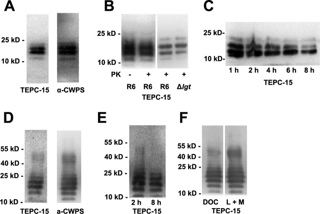
FIG 1.
Western blot analysis of S. pneumoniae TAs. (A) SDS-soluble TA was separated by 16% Tricine SDS-PAGE and probed with the antibodies indicated. TEPC-15 is a mouse IgA monoclonal antibody specific for P-Cho; α-CWPS is a rabbit CWPS (structurally, a PG-TA complex of pneumococci) polyclonal antiserum. (B) The banding pattern of SDS-soluble TAs was not influenced by proteinase K digestion and lgt mutation in strain R6, showing that neither proteinase K nor lgt mutation impaired the TA banding pattern. (C) Banding pattern of SDS-soluble TAs from strain R6 following digestion with mutanolysin plus lysozyme for the times indicated, showing reduced TA banding intensity with increasing digestion times. (D) Banding pattern of TAs from whole R6 bacteria digested with mutanolysin plus lysozyme for 2 h and probed with the antibodies indicated. (E) Banding pattern of TAs from whole R6 bacteria digested with mutanolysin plus lysozyme for the times indicated. (F) Banding pattern of TAs from whole R6 bacteria treated with DOC or lysozyme plus mutanolysin (L + M). The positions of size markers (molecular masses in kilodaltons are shown) are indicated.

