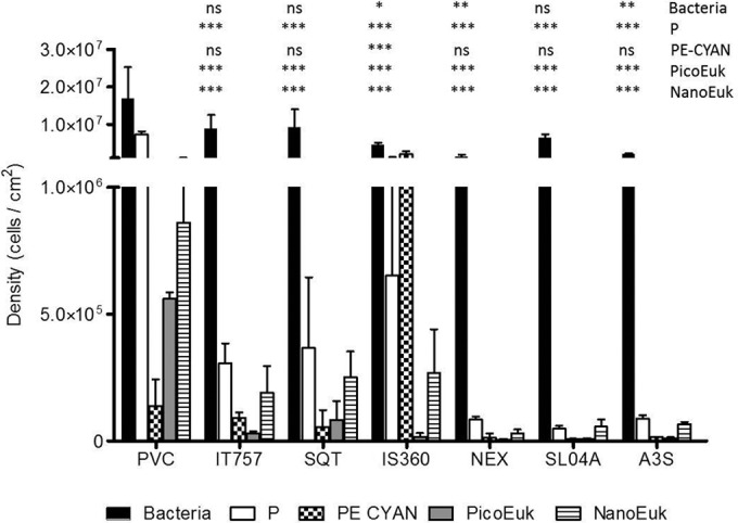
FIG 3.

Microorganism density (cells/cm2) on the seven coatings, determined by using flow cytometry. Data for NanoEuk, PicoEuk, low-pigmented picoorganisms (P), PE-CYAN, and bacteria are shown. * (P < 0.05), ** (P < 0.01), and *** (P < 0.001) indicate that the density of microorganisms was significantly different on the coating compared to that on PVC. ns, not significant.
