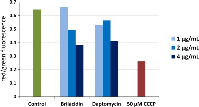
FIG 2.
Membrane depolarization of Staphylococcus aureus. Newman cultures suspended in buffer supplemented with Ca2+ were treated with 1, 2, and 4 μg/ml of brilacidin and daptomycin for 30 min and stained with DiOC2(3) for 15 min. Red/green fluorescence emission ratios were determined for 30,000 cells by flow cytometry, with a decrease in the red/green ratio indicating depolarization of the membrane. The protonophore CCCP was used as a positive control for membrane depolarization.

