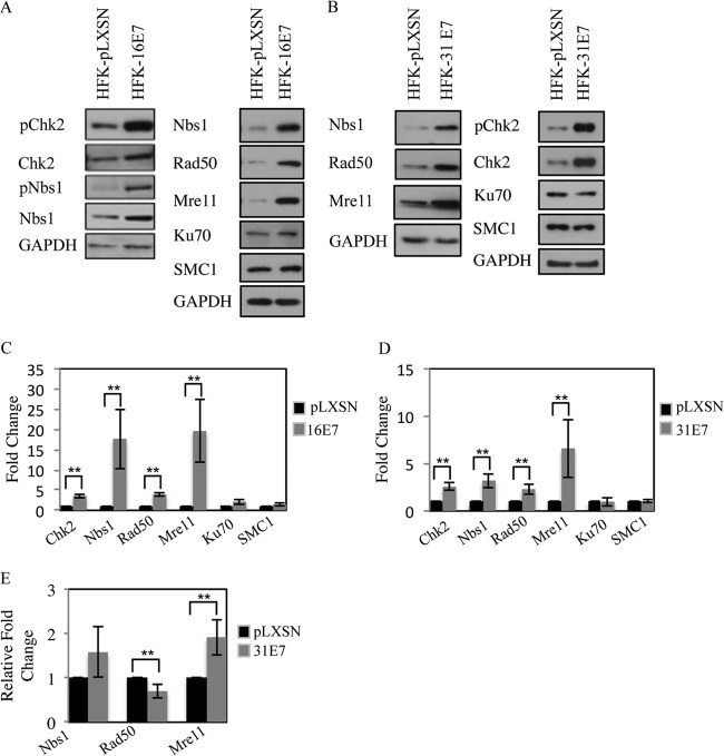
FIG 1.
Expression of HPV E7 increases levels of proteins associated with detection and repair of DNA damage. (A) Whole-cell extracts were harvested from HFKs stably expressing pLXSN vector control or pLXSN-HPV16 E7. Immunoblotting was performed using antibodies to phosphorylated Chk2 (Thr68) (pChk2), total Chk2, phosphorylated Nbs1 S343 (pNbs1), total Nbs1, Rad50, Ku70, and SMC1. (B) Lysates harvested from HFK-pLXSN or HFK-pLXSN HPV31 E7 cells were analyzed by Western blotting using antibodies to pChk2, Chk2, Nbs1, Rad50, Mre11, Ku70, and SMC1. GAPDH served as a loading control. (C and D) Bar graphs demonstrate the average expression level of target proteins, normalized to GAPDH, in at least four independent Western blot analyses, including data from panels A and B. Densitometry was performed using ImageJ software. The statistical analysis was assayed by 2-tailed t test. Data are means ± standard errors. **, P value less than 0.05. (E) Quantitative real-time PCR of gene expression analysis in HFK-pLXSN and HFK-pLXSN-31E7 lines. Expression levels are shown relative to HFK-pLXSN cells and were calculated using GAPDH as a reference gene. Shown is the relative fold change in gene expression over three independent experiments. The statistical analysis was assayed by 2-tailed t test. Data are means ± standard errors. **, P value less than 0.05.

