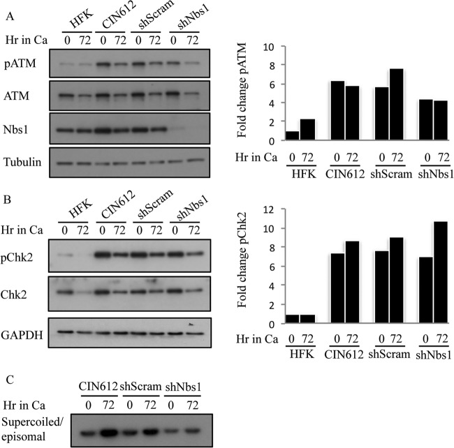
FIG 11.
Phosphorylation of ATM and Chk2 is maintained with Nbs1 knockdown upon differentiation. (A) Whole-cell lysates were harvested from HFKs and CIN612 9E cells, as well as CIN612 9E cells stably expressing shScramble or shNBS1 cells at T0 and 72 h after differentiation in high-calcium medium. Immunoblotting was performed using antibodies to phosphorylated ATM (Ser1981) (pATM), total ATM, and Nbs1. Tubulin was used as a loading control. Protein levels were quantified using ImageJ, with phosphorylated protein levels normalized first to total levels and then to tubulin. Levels for this representative experiment are graphed as fold change compared to the T0 HFK sample, which is set to 1. (B) Whole-cell lysates were harvested from HFK, CIN612 9E, CIN612 9E shScramble, and CIN612 9E shNBS1 cells at T0 and 72 h after differentiation in high-calcium medium. Immunoblotting was performed using antibodies to phosphorylated Chk2 (Thr68) (pChk2), total Chk2, and Nbs1. GAPDH was used as a loading control. Protein levels were quantified using ImageJ as indicated above. Shown is a representative experiment where levels are graphed as fold change compared to the T0 HFK sample, which is set at 1. (C) DNA was harvested from CIN612 9E, CIN612 9E shScramble, and CIN612 9E shNBS1 cells at T0 and after 72 h of differentiation in high-calcium medium and linearized by digestion with HindIII. HPV episomes were visualized via Southern blot analysis. Results shown are representative observations of four or more independent experiments.

