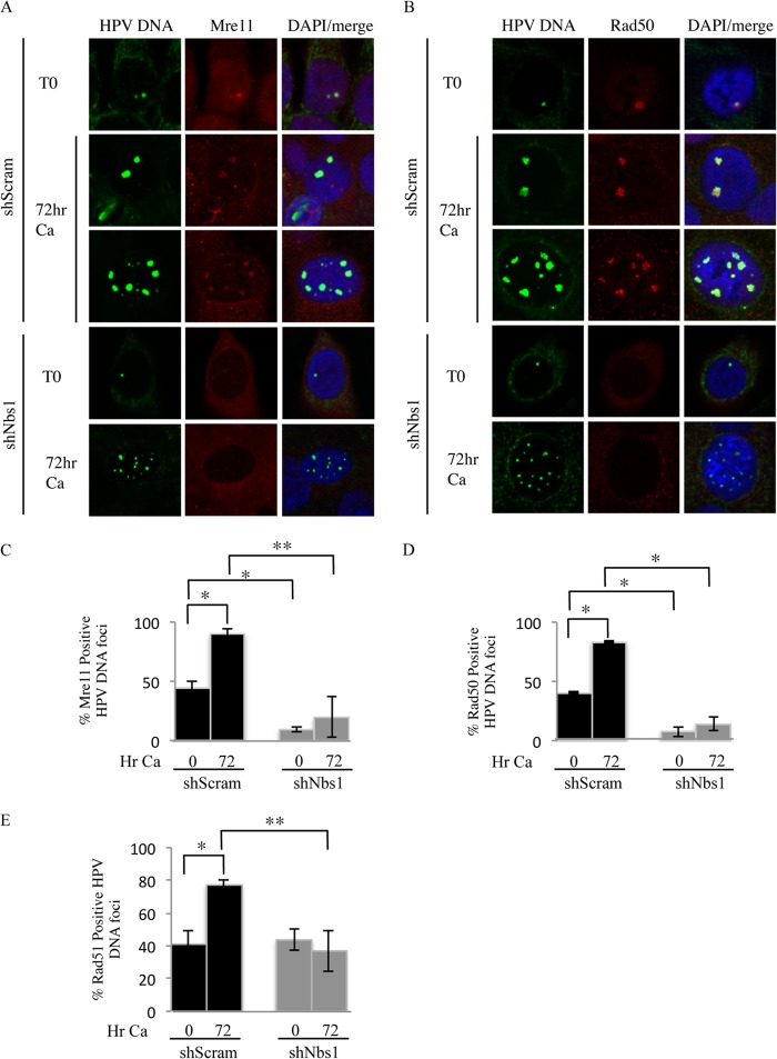
FIG 9.
Nbs1 knockdown results in decreased localization of Mre11, Rad50, and Rad51 to viral genomes. (A and B) Immunofluorescence (IF) for Mre11 (red) (A) and Rad50 (red) (B), followed by fluorescence in situ hybridization (FISH) for HPV DNA (green), was performed on CIN612 9E cells stably maintaining the Scramble or Nbs1 shRNAs at T0 (undifferentiated), as well as after 72 h of differentiation in high-calcium medium. Cellular DNA was counterstained with DAPI. (C and D) Bar graphs represents quantification of percentages of HPV DNA focus-positive cells that were also positive for Mre11 (C) and Rad50 (D). (E) Quantification of percentage of cells positive for HPV DNA foci by FISH that were also positive for Rad51 by IF in CIN612 9E-shScram and -shNbs1 cells at T0, as well as after 72 h of differentiation in high-calcium medium. The data represent the averages of at least three independent experiments. The statistical analysis was assayed by 2-tailed t test. Data are means ± standard errors. **, P value less than 0.05; *, P value less than 0.01; Ca, calcium.

