
FIG 3.
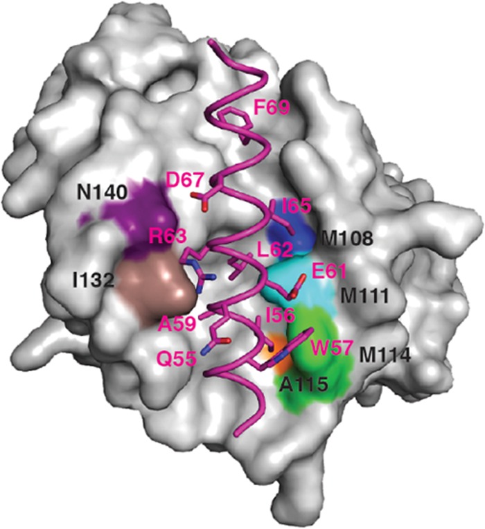
Structure of F1L in complex with the Bim BH3 peptide highlighting the F1L binding pocket hydrophobic residues. Shown is the molecular surface of F1L in complex with the BH3 peptide of BimL (in magenta). The FIL binding pocket residues that facilitate hydrophobic contact with specific amino acids of BimL are highlighted in colors. Included are F1L(N140), which is purple; F1L(I132), which is brown; F1L(A119W), which is orange; F1L(M114), which is green; F1L(M111), which is aqua; and F1L(M108), which is blue.