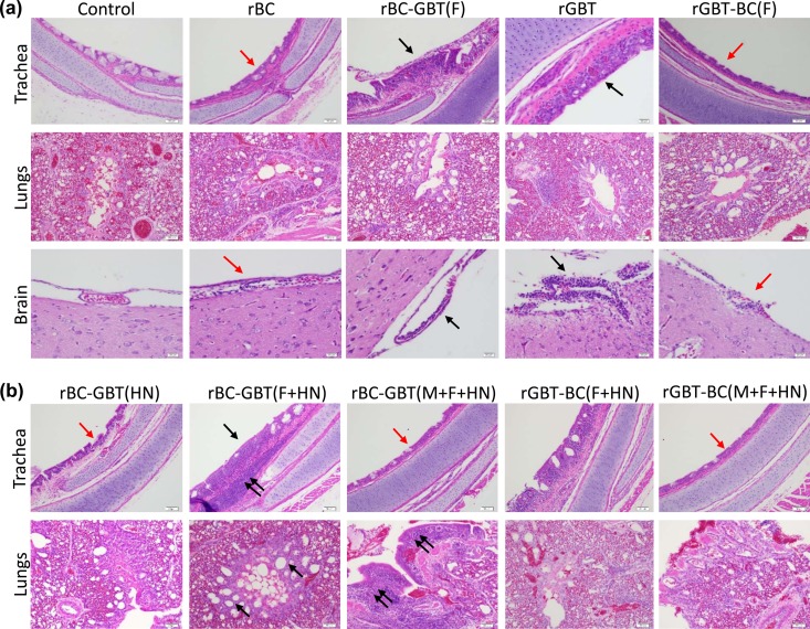
FIG 8.
Histopathology of trachea, lungs, and brain tissues of 2-week-old chickens infected oculonasally with parental and chimeras of NDV strains BC and GBT after swapping of the envelope-associated protein genes. Birds were sacrificed at 3 dpi, and tissue was fixed with formalin, sectioned, and stained with hematoxylin and eosin. (a, trachea) The rGBT parent caused severe inflammation of the tracheal mucosa (black arrows), which also was observed with rBC-GBT(F), whereas the rBC parent and rGBT-BC(F) caused attenuation of the tracheal epithelium (red arrows) with minimal inflammation. (a, lungs) There was no significant difference observed among rBC and rGBT and their F gene swap constructs. (a, brain) Strain GBT caused substantial lymphoplasmacytic infiltration in the meninges (black arrows), which also was observed with rBC-GBT(F), whereas rBC and rGBT-BC(F) were associated with reduced lymphoplasmacytic infiltration (red arrows). (b, trachea) rBC-GBT(F+HN) caused severe inflammation of the tracheal mucosa (black arrow) and submucosa (double black arrows) compared to mucosal attenuation in the rBC-GBT(HN), rBC-GBT(M+F+HN), and rGBT-BC(M+F+HN) groups (red arrows) but the rGBT-BC(F+HN) group showed moderate mucosal inflammation. (b, lungs) rBC-GBT(F+HN) caused hyperplasia of the parabronchi (black arrows), and rBC-GBT(M+F+HN) caused severe parabronchial inflammation (red arrows), whereas this was minimal with rBC-GBT(HN), rGBT-BC(F+HN), and rGBT-BC(M+F+HN). In these images it is evident that when GBT F was exchanged with that of BC, this increased the virulence of BC alone or in combination with GBT HN. In contrast, when BC F was exchanged with that of GBT it reduced the virulence of GBT.

