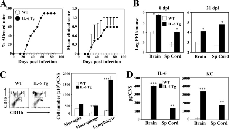
FIG 2.
B6 IL-6 Tg mice infected with TMEV develop demyelinating disease and exhibit elevated viral load. (A) Development of demyelinating disease in TMEV-infected WT (n = 10) and IL-6 Tg (n = 10) mice was monitored. (B) Viral loads in the CNS (n = 3) of infected mice were determined using plaque assays at 8 and 21 days postinfection. (C) Flow cytometry of CNS-infiltrating cells bearing CD45 and CD11b in WT (n = 3) and IL-6 Tg mice (n = 3) at 8 days after virus infection. The bar graph shows the number of CD45int CD11b+ microglia, CD45hi CD11b+ macrophages, and CD45hi CD11− lymphocytes in the CNS of infected mice. (D) Levels of IL-6 and KC in the CNS of virus-infected WT B6 and IL-6 Tg mice were assessed using specific ELISA at 8 days postinfection. Sp Cord, spinal cord.

