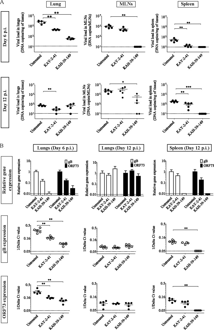
FIG 5.
Analysis of MHV-68 infection in different organs of untreated and treated mice at the acute and latent stages. (A) Viral DNA copies were quantified in the lungs, mediastinal lymph nodes (MLNs), and spleens of intranasally infected mice (104 PFU of MHV-68) and treated intraperitoneally with KAY-2-41 and KAH-39-149 for five consecutive days. Each group contained five mice. The values are given as the mean log viral copy number per mg of tissue ± standard deviation (SD). The dashed line represents the limit of detection set on 10 copies of viral DNA. (B) The upper panels represent the level of gB (white bars) and ORF73 (black bars) expression relative to those of the untreated control and normalized to the endogenous control glyceraldehyde-3-phosphate dehydrogenase (GAPDH). The error bars display the calculated maximum (RQMax) and minimum (RQMin) expression levels that are equivalent to the standard error of the mean expression level (RQ value). The RQMax is represented as ΔΔCT + s and the RQMin as ΔΔCT − s, where s is the SD of the ΔΔCT value. The lower panels represent the 1/delta CT values obtained from each mouse in the untreated and treated groups, and on which the Mann-Whitney U tests were done to compare the untreated and treated groups: *, P < 0.05; **, P < 0.01; ***, P < 0.001. The dashed line represents the limit of detection.

