


FIG 4.
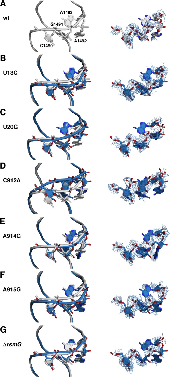
Backbone shifts in the decoding site. (Left) Wild-type and mutant structures were aligned using the phosphate atoms of 16S rRNA residues 50 to 500. Gray, wild type; pale cyan, mutants. Side chains for residues C1490, G1491, A1492, and A1493 are also shown. (Right) Composite omit 2mFo − DFc electron density maps for the same four residues. (A) Wild type; (B) wild type and U13C; (C) wild type and U20G; (D) wild type and C912A; (E) wild type and A914G; (F) wild type and A915G; (G) wild type and ΔrsmG.