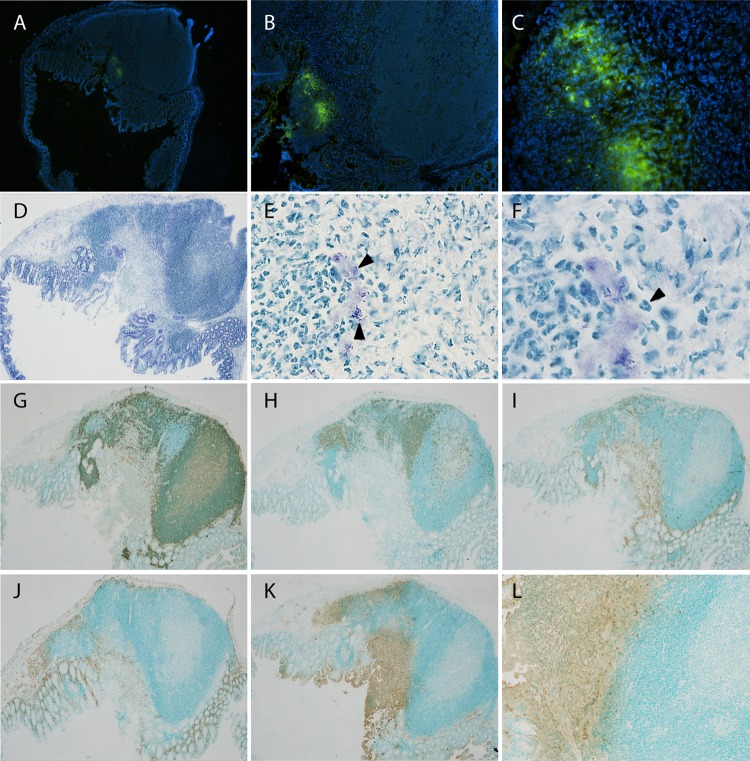
FIG 5.
Characterization of cecal aggregates colonized by Y. pseudotuberculosis. Cecal tissues from mice positive for bioluminescent signals at day 34 postinfection were sectioned and analyzed by immunofluorescence and immunohistochemistry. (A to C) Immunofluorescent staining using anti-Yersinia polyclonal serum, detected by anti-rabbit-Al488 (green). Nuclei were stained with DAPI (blue). Magnifications: ×4 (A), ×10 (B), and ×40 (C). (D to F) Toluidine blue-stained sections. Magnifications: ×4 (D), ×60 (E), and ×100 (F). Arrowheads in panel E indicate bacteria, and that in panel F indicates a polymorphonuclear cell. (G to L) Immunohistochemical staining using monoclonal antibodies with amplification of signal by streptavidin-biotin and horseradish peroxidase-conjugated antibiotin: anti-B220 for B cells (G), anti-CD3 for T cells (H), anti-CD11c for dendritic cells (I), anti-F4/80 for macrophages (J), and anti-Ly6G/6C for PMNs (K and L). Positive cells are brown (diaminobenzidine), and the background is green (methyl green). Magnifications: ×4 (G to K) and ×10 (L). The stainings shown are representative from 6 individual persistent infected ceca.

