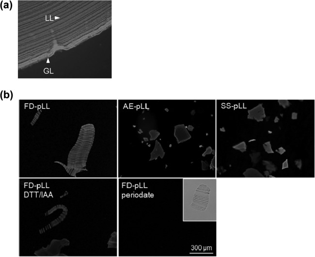
FIG 1.
The LL tends to give rise to flat particles upon disruption. Shown are fluorescence microscopy images of the native LL (a paraffin-embedded section from a natural cattle infection) (a) and pLL preparations (b). The images are of pLL prepared by the three methods explained in the text (FD-, EA-, and SS-pLL); note that the laminations present in the native LL are observed only in FD-pLL. Also shown are FD-pLL preparations subjected to reduction/alkylation of disulfides (DTT/IAA) or oxidation of carbohydrate residues with periodate (followed by reduction of aldehydes with borohydride). In both panels, the samples have been labeled with the lectin PNA conjugated to FITC for easier visualization. Periodate oxidation abrogates PNA binding, as expected, but the physical preparation of the material does not change, as can be observed in the clear-field image (inset). The pLL preparations shown are of bovine host origin, except for SS-pLL, which is from a mouse host.

