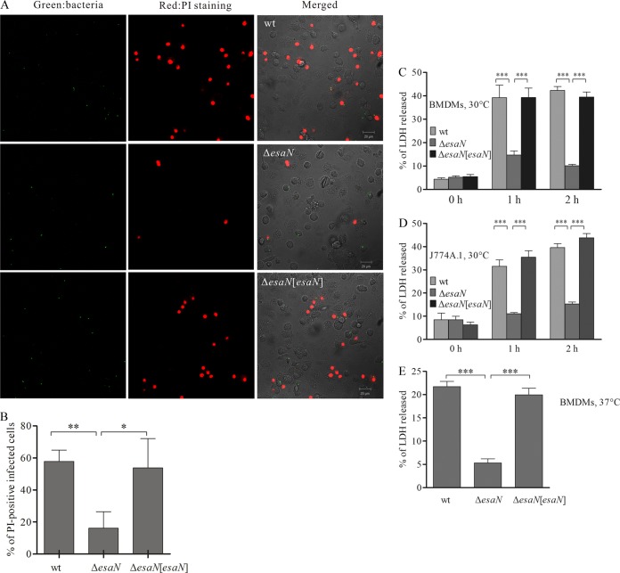
FIG 2.
T3SS-dependent cytotoxicity in BMDMs and J774A.1 cells. (A) Confocal micrographs of PI uptake by BMDMs. BMDMs were infected with GFP-expressing strains for 2 h and stained with PI in Opti-MEM without fixation. Green, bacteria; red, PI-positive BMDMs. Bar, 50 μm. (B) Quantification of PI-positive infected cells. More than 300 infected cells for each infection were counted. (C) T3SS-dependent cytotoxicity of E. tarda PPD130/91 in BMDMs at 30°C determined by LDH assay. BMDMs were infected with different bacterial strains, and the culture supernatants were collected and processed for the LDH release assay. (D) T3SS dependent cytotoxicity of E. tarda PPD130/91 in J774A.1 at 30°C. (E) T3SS-dependent cytotoxicity of E. tarda PPD130/91 in BMDMs at 37°C. Samples were collected 1 h after uptake for analysis. Means ± SEM from three experiments are shown. *, P < 0.05; **, P < 0.01; ***, P < 0.001.

