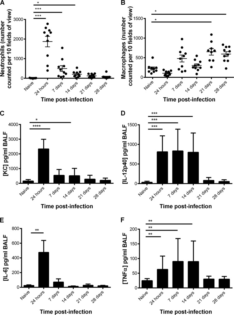
FIG 3.
Inflammatory cells and cytokines in BALF collected from CBA/Ca mice after intranasal infection with S. pneumoniae strain LgSt215 (n = 10). (A) Number of macrophages; (B) number of neutrophils. Cells were counted in 10 fields of view at ×400 magnification. C, D, E, and F show the levels of the cytokines KC, IL-12 p40, IL-6, and TNF-α, respectively. Mann-Whitney nonparametric t tests were used to compare differences between pairs of time points: *, P values of <0.05 and >0.01; **, P values of <0.01 and >0.001; ***, P values of <0.001 and >0.0001; ****, P values of <0.0001.

