
FIG 4.
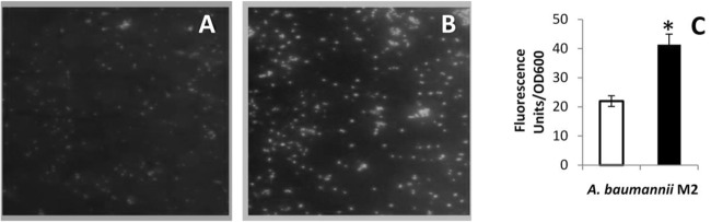
(A and B) Representative fluorescence microscope images of CM-H2DCFDA-stained A. baumannii M2 grown in the absence (A) and presence of pyocyanin (B). Magnification, ×40. (C) Quantitation of fluorescence in A. baumannii M2 grown in the absence (□) and presence (■) of pyocyanin. Data are means from three experiments. *, P ≤ 0.001.