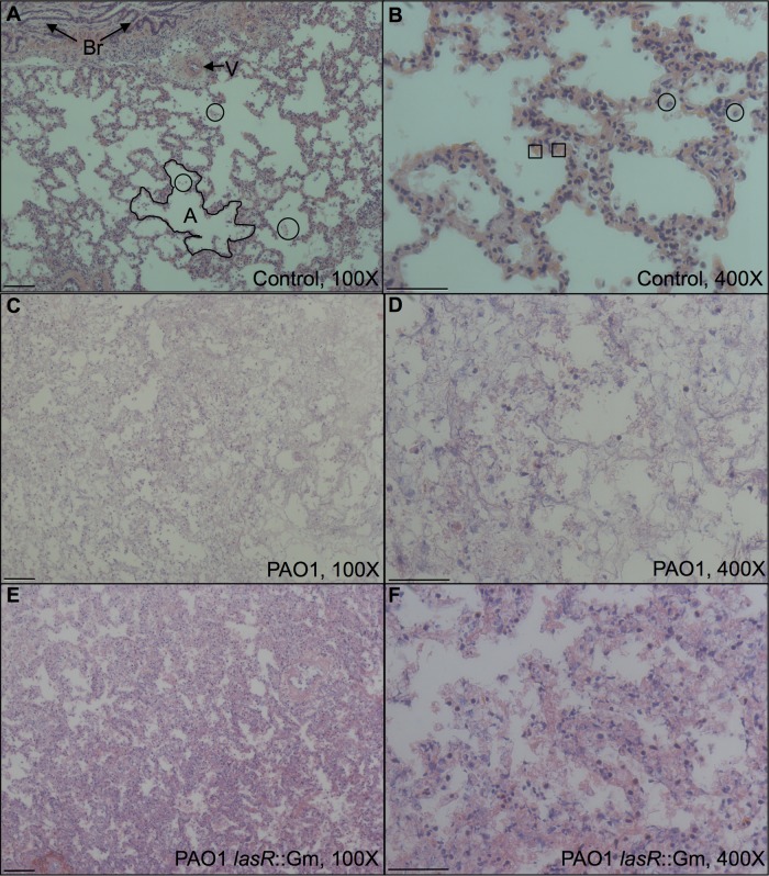
FIG 2.
Micrographs of tissue after 24 h in ASM, fixed and stained with H&E, which colors nuclei dark blue and other structures (cytoplasm, collagen, etc.) pink. (A and B) Mock-infected control; (C and D) infected with WT P. aeruginosa; (E and F) infected with the lasR::Gm mutant. Panels A, C, and E show tissue at magnification ×100 with a 100 μM scale bar; panels B, D, and F show tissue at magnification ×400 with a 50 μM scale bar. In panel A, note two bronchioles (Br) with diagnostic folded epithelium of brush border, example of a blood vessel (V), and lace-like pattern of alveoli defined by thin epithelium (example outlined; A). Small patches of cellular debris are visible in the alveoli (three examples are circled). In panel B, occasional cells with horseshoe-shaped nuclei (circled) are visible, which may represent neutrophils, along with enucleate red blood cells (two examples are boxed). Note in panels C and D the loss of clear epithelium, lower number of nuclei, and a decreased volume of airspace. In panels E and F, this change is less extreme, with thickened outlines of epithelium still discernible.

