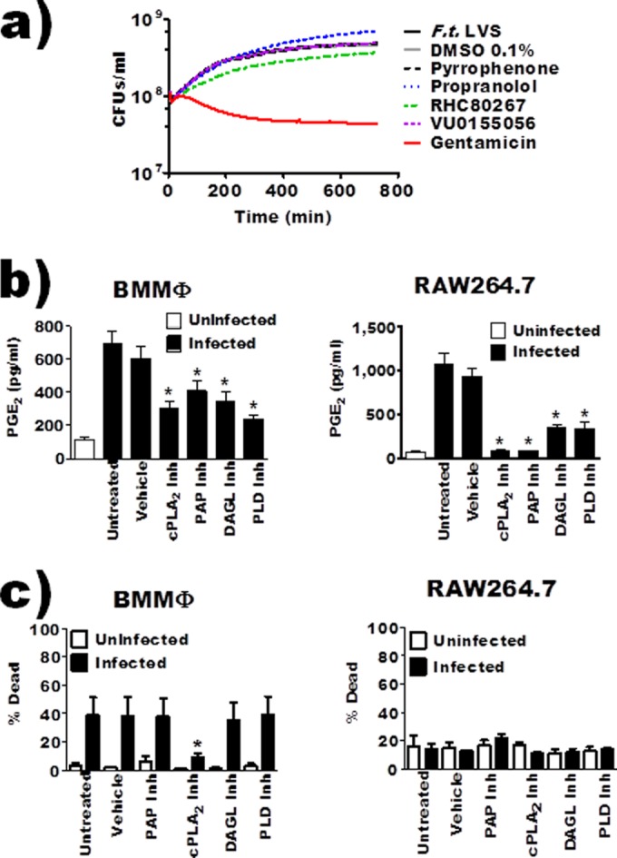
FIG 3.

Inhibition of macrophage cPLA2 (pyrrophenone), PAP (propranolol), DAGL (RHC80267), or PLD (VU0155056) reduced F. tularensis-induced prostaglandin E2 biosynthesis by infected macrophages. (a) F. tularensis LVS was inoculated in broth medium and left untreated or incubated with inhibitors/vehicle and allowed to grow over 12 hours. Bacterial growth was measured as a function of absorbance at 600 nm. Curves represent average growth from 3 independent experiments. (b) BMDMs or RAW 264.7 macrophages were pretreated with inhibitors and then inoculated with F. tularensis LVS at an MOI of 200:1; PGE2 in the supernatant was quantified by enzyme-linked immunosorbent assay 24 hours after inoculation. Data represent means ± SEMs. n = 9 (BMDMs); n = 17 (RAW cells). (c) LIVE/DEAD Fixable Yellow stain analysis was performed to verify that inhibitors were not causing cell death or increased cell death during F. tularensis infection. BMDMs or RAW 264.7 macrophages were incubated with vehicle or inhibitor for 20 hours at 37°C (n = 6). Asterisks denote a statistical difference compared to the untreated infected group (P ≤ 0.05). Graphs are means of pooled experiments ± SEMs.
