FIG 3.
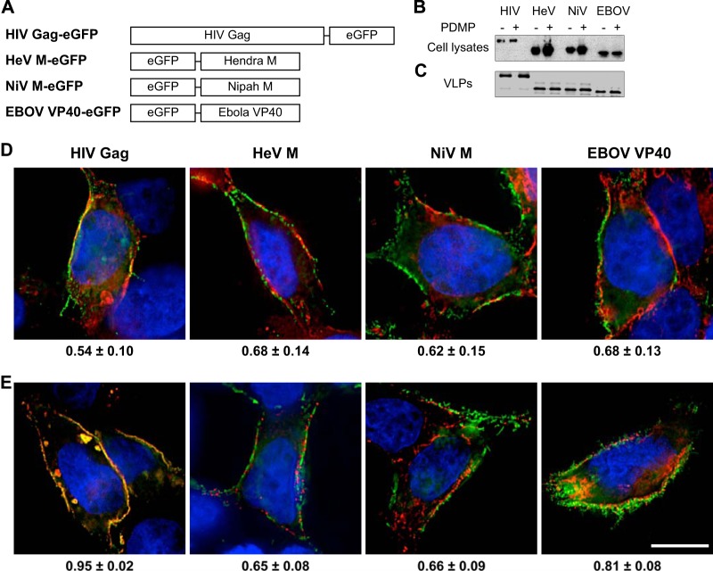
Paramyxovirus and filovirus VLPs bud from GSL-enriched lipid microdomains at the plasma membrane. (A) Constructions of HeV M, NiV M, and EBOV VP40 fusion proteins. (B and C) Western blot analysis of eGFP fusion proteins in transfected HEK293T cell lysates (B) and VLPs in the supernatants (C) in the presence or absence of PDMP. (D) Localization of eGFP fusion proteins in transfected HEK293T cells stained with CTxB for GM1 (red) and with DAPI for the nucleus (blue). The values at the bottom are Pearson's colocalization coefficients of the means ± SDs. (E) Localization of Gag-mCherry and the indicated eGFP fusion proteins in transfected HEK293T cells stained with DAPI (blue). The values at the bottom are Pearson's colocalization coefficients of the means ± SDs. Bar, 10 μm.

