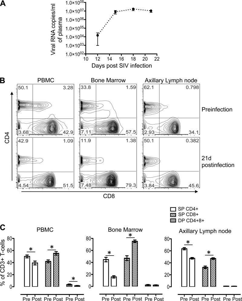
FIG 1.
(A) Mean (± standard errors) plasma viral loads in macaques during the acute phase of infection with SIVMAC251, as determined by RT-PCR (n = 9). (B) Representative contour plots showing SP CD4+ or CD8+ and DP CD4+ CD8+ T-cell populations in PB, BM, and ALN mononuclear cells before and 21 days after SIVMAC251 infection. (C) Mean percentages (± standard errors) of SP CD4+ or CD8+ and DP CD4+ CD8+ T cells of SIV-infected macaques before infection (Pre) and 21 days after infection (Post) (n = 9). Plots were generated by gating CD3+ T cells. Asterisks indicate statistically significant differences from the preinfection levels in the respective cell populations (P < 0.05).

