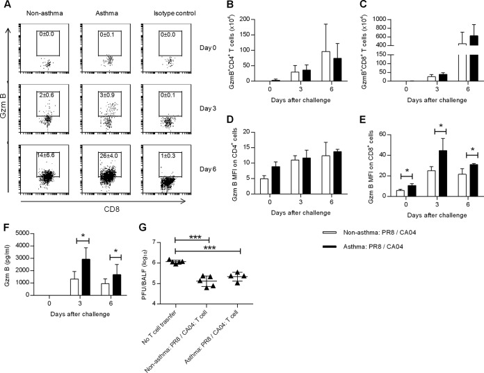
FIG 6.
Antiviral T-cell effector function is not compromised in asthmatic mice. (A) Representative dot plots showing the mean percentages ± SD of gzmB+ CD8+ T cells on days 0, 3, and 6 following CA04 challenge (3 or 4 mice/group). Numbers of gzmB+ CD4+ (B) and gzmB+ CD8+ (C) pulmonary T cells in nonasthmatic:PR8/CA04 and asthmatic:PR8/CA04 mice as assessed by intracellular flow cytometry on days 0, 3, and 6 post-CA04 challenge (3 or 4 mice/group). MFI of gzmB+ CD4+ (D) and CD8+ (E) lung T cells (3 or 4 mice/group). (F) gzmB levels in BALF on days 0, 3, and 6 post-CA04 infection (7 or 8 mice/group from two independent experiments). (G) Five days after CA04 challenge, CD3+ T cells were isolated from the lungs of nonasthmatic:PR8/CA04 and asthmatic:PR8/CA04 mice and transferred into day 2 CA04 (5,000 PFU)-infected recipients. Viral lung titers in the recipients 5 days after T-cell transfer are shown (4 or 5 mice/group). *, P < 0.05; ***, P < 0.001.

