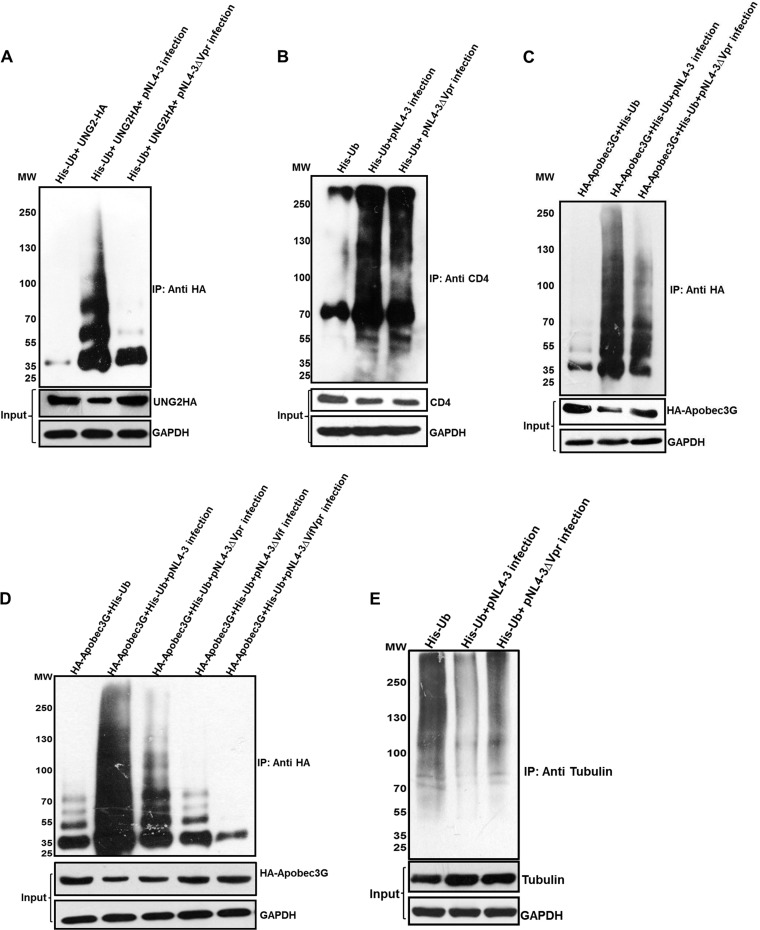
FIG 5.
Ubiquitination and degradation of different proteins. (A) HEK 293T cells were cotransfected with HA-UNG2 and His-Ub. After 12 h, the cells were infected with equal multiplicities of infection of pNL4-3/pNL4-3Δvpr VSV-G-pseudotyped virus. After 36 h, MG132 treatment was given for 8 h, and ubiquitinated proteins were enriched by using Ni-NTA beads. Immunoblot analysis was done by using an anti-HA antibody. (B) TZM-bl cells were transfected with His-Ub. After 12 h, the cells were infected with equal multiplicities of infection of pNL4-3/pNL4-3Δvpr VSV-G-pseudotyped virus. After 36 h, MG132 treatment was given for 8 h, and ubiquitinated proteins were enriched by using Ni-NTA beads. Immunoblot analysis was done by using anti-CD4 antibody. (C) HEK 293T cells were cotransfected with HA-APOBEC3G and His-Ub. After 12 h, the cells were infected with equal multiplicities of infection of pNL4-3/pNL4-3Δvpr VSV-G-pseudotyped virus. After 36 h, MG132 treatment was given for 8 h, and ubiquitinated proteins were enriched by using Ni-NTA beads. Immunoblot analysis was done by using an anti-HA antibody. (D) HEK 293T cells were cotransfected with HA-APOBEC3G and His-Ub. After 12 h, the cells were infected with equal multiplicities of infection of pNL4-3/pNL4-3Δvpr/pNL4-3Δvif/pNL4-3ΔvifΔvpr VSV-G-pseudotyped virus. After 36 h, MG132 treatment was given for 8 h, and ubiquitinated proteins were enriched by using Ni-NTA beads. Immunoblot analysis was done by using an anti-HA antibody. (E) HEK 293T cells were transfected with His-Ub. After 12 h, the cells were infected with equal multiplicities of infection of pNL4-3/pNL4-3Δvpr VSV-G-pseudotyped virus. After 36 h, MG132 treatment was given for 8 h, and ubiquitinated proteins were enriched by using Ni-NTA beads. Immunoblot analysis was done by using an anti-tubulin antibody. Levels of proteins are shown as the input (without MG132 treatment). GAPDH was used as a loading control.

