FIG 2.
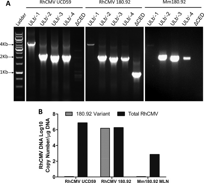
Conventional and real-time PCR amplification of RhCMV-specific segments within the UL/b′ region. DNA extracted from RhCMV UCD59 virus stock, RhCMV 180.92 virus stock, and frozen mesenteric lymph node (MLN) tissue collected at necropsy from the SIV-infected rhesus macaque Mm180.92, the monkey from whom RhCMV 180.92 was originally recovered, were used. (A) Conventional PCR runs with UL/b′ region primers on RhCMV UCD59 (left panel), RhCMV 180.92 (middle panel), and MLN from Mm180.92 (right panel). Segments within the UL/b′ region corresponding to the low-passage-number WT-like isolate RhCMV UCD59 (ULb′-1 to -4) are present in RhCMV strain 180.92 (middle panel). Amplicon specific to the truncated sequence of RhCMV 180.92 (ΔCED) is minimal to completely absent from DNA isolated from RhCMV UCD59 (left panel) and animal Mm180.92 (right panel), respectively. (B) Quantitative real-time PCR showing total and 180.92 truncated variant-specific RhCMV copy numbers per μg of DNA extracted from RhCMV UCD59, RhCMV 180.92, and MLN of Mm180.92. Median values of three replicate wells shown.

