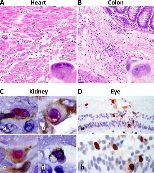
FIG 3.
Histopathologic analysis of CMV localization in tissues of a SIV-infected CMV-seronegative rhesus macaque experimentally inoculated with RhCMV 180.92. Photomicrographs of H&E-stained and immunohistochemically stained tissue sections from a rhesus macaque with simian AIDS and disseminated CMV after SIV and RhCMV 180.92 coinfection. (A and B) RhCMV-induced inflammation in the heart (A) and colon (B) characterized by mild edema and tissue infiltration by lymphocytes and histiocytes admixed with cytomegalic and karyomegalic cells containing multiple large round to oval magenta-colored CMV intranuclear inclusion bodies surrounded by clear halo and chromatin margination (magnification, ×200; inset magnification, ×600). (C) Immunohistochemical analysis of kidney tissue showing distribution of RhCMV 180.92 infection in a wide range of cell types identified by double immunostaining of CMV-IE1 (magenta) with CD31 for endothelial cell identification (brown [a]), cytokeratin for epithelial cell identification (brown [b]), vimentin for fibroblast identification (brown [c]), and CD68 for macrophage identification (brown [d]). Magnification, ×600. (D) RhCMV-induced retinitis characterized by the presence of numerous immunohistochemically positive cells for CMV-IE1 (brown) associated with focal retinal degeneration and disruption of retinal internal and external nuclear layers shown at low (×200 [a]) and high (×400 [b]) magnifications.

