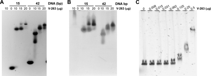
FIG 3.

Protein V-DNA binding assays. (A) An electrophoretic mobility shift assay (EMSA) was performed with 2 μg of 15 or 42 bp dsDNA that was incubated with the indicated amounts of recombinant protein V at 25°C for 20 min prior to analysis on an 0.8% agarose gel and then stained with SYBR green to visualize the DNA. (B) The same gel was stained with Simply Blue to visualize protein. (C) EMSA was performed with 150 ng of linearized bacmid (39 kbp) that was incubated with the indicated amounts of protein V at 25°C for 20 min prior to analysis on a 0.8% agarose gel and then stained with SYBR green.
